Quick Answer
Research on red light therapy and collagen synthesis suggests that specific light treatments can influence collagen-related activity in skin. Human studies have reported changes in collagen fibers and some wrinkle measurements. Experimental studies also identify signaling pathways involved in collagen production and preservation, although those mechanisms do not guarantee the same results from every home device.
Key Takeaway
Red light therapy and collagen synthesis involve more than producing a structural protein. Fibroblast signaling, collagen breakdown, and the organization of the surrounding tissue all matter. Understanding what researchers measured helps you judge claims about firmer skin and fewer wrinkles.
Fine lines may show up on the surface, but the processes that shape them run deeper. Beneath your skin’s outer layer, fibroblasts help build and maintain the collagen-rich network that gives tissue strength. Research on red light therapy and collagen synthesis explores whether light can influence the molecular signals directing that work, and how those changes might translate into smoother, firmer-looking skin.
The science involves gene transcription, cellular signaling, and extracellular matrix remodeling. These terms describe how cells receive instructions, produce structural proteins, and maintain the material around them. Understanding those steps helps you make sense of what the research actually shows.
What does collagen synthesis actually mean?
Collagen synthesis is the production of collagen, a family of structural proteins. In the dermis, the layer beneath your skin’s outer surface, collagen fibers help provide strength.
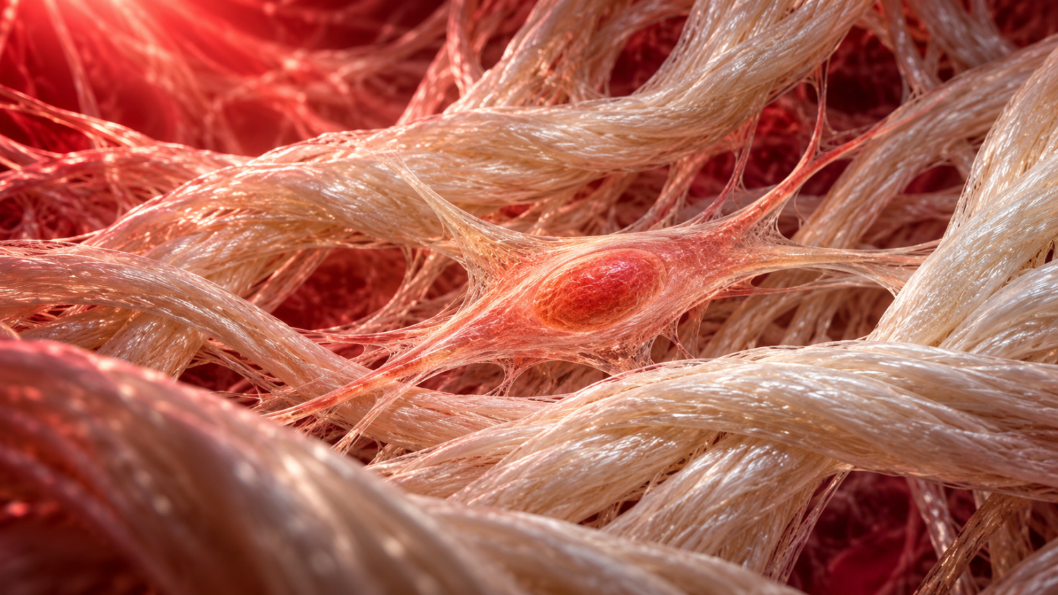
Dermal fibroblasts produce collagen and other components of the extracellular matrix. This matrix is the material surrounding cells and helping organize tissue.
Production involves several stages. Cells express collagen genes, assemble precursor molecules called procollagen, and release them. Further processing allows collagen molecules to assemble into fibrils and larger fibers.
Think of construction materials moving from an order to a finished structure. Increasing orders does not tell you how much usable structure was completed.
That distinction matters when reading about red light therapy and collagen synthesis. A rise in collagen-related messenger RNA shows a change in gene expression. It does not, by itself, prove that mature collagen fibers increased or wrinkles improved.
Why aging changes the fibroblast environment
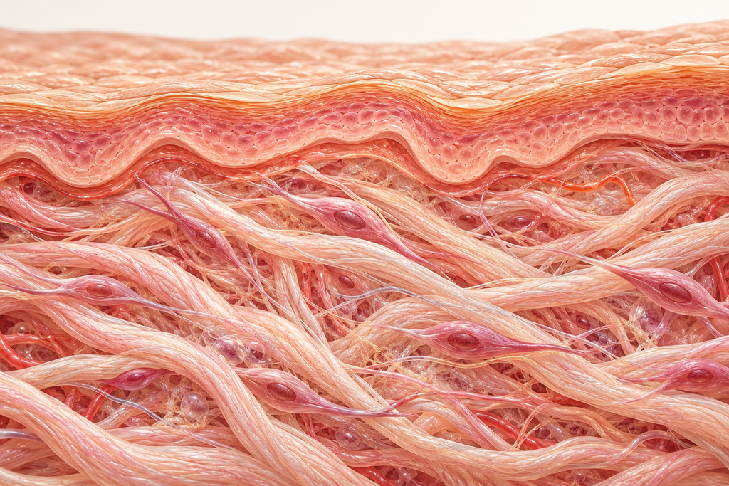
Fibroblasts respond to their surroundings. Their contact with the extracellular matrix provides mechanical signals that influence their behavior.
In a study of aged human skin, researchers linked fragmented collagen with reduced fibroblast stretch, oxidative stress, and increased expression of matrix metalloproteinase-1, or MMP-1. This enzyme helps break down collagen.
Separate human skin and cell research connected reduced fibroblast size and mechanical force with weaker TGF-β receptor signaling and reduced production of important matrix components.
In plain language, a deteriorating support structure can also change how its maintenance cells work.
This helps explain why red light therapy and collagen synthesis belong within a broader discussion of skin remodeling. The cells, signals, and surrounding fibers influence one another.
How might light influence fibroblast signaling?
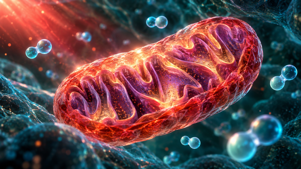
Photobiomodulation, or PBM, uses light to influence biological activity. A photochemical response begins when a molecule absorbs light.
Researchers investigate how this can affect cellular metabolism and signaling. In cultured skin fibroblasts, specific light exposures have been studied for effects on mitochondrial membrane potential and redox balance. pmc.ncbi.nlm.nih.gov
Membrane potential is an electrical difference across a membrane. Redox balance concerns linked oxidation and reduction reactions, which involve electron transfer.
Your cells use these processes for both function and communication. Our guide to mitochondrial redox signaling explores that chemistry in more detail.
For red light therapy and collagen synthesis, the next question is whether initial cellular changes influence downstream protein production. Measuring a mitochondrial response alone cannot answer that.
TGF-β/SMAD signaling and collagen gene transcription
One advanced pathway under investigation is TGF-β/SMAD signaling. TGF-β stands for transforming growth factor beta, a signaling protein involved in tissue regulation.
When it binds to receptors, it can activate intracellular messengers called SMAD proteins. Phosphorylation means adding a phosphate group, a chemical modification that can change a protein’s activity. Nuclear translocation means movement into the nucleus, where genes are regulated.
A 2025 study in naturally aging mice and mouse fibroblasts linked red light exposure with TGF-β/SMAD activity and increased type I collagen transcription. Blocking the pathway reduced the response.
These experiments provide a possible mechanism for red light therapy and collagen synthesis. They do not establish that every consumer device activates the same pathway in human facial skin.
AKT/Nrf2/HO-1 signaling and collagen preservation
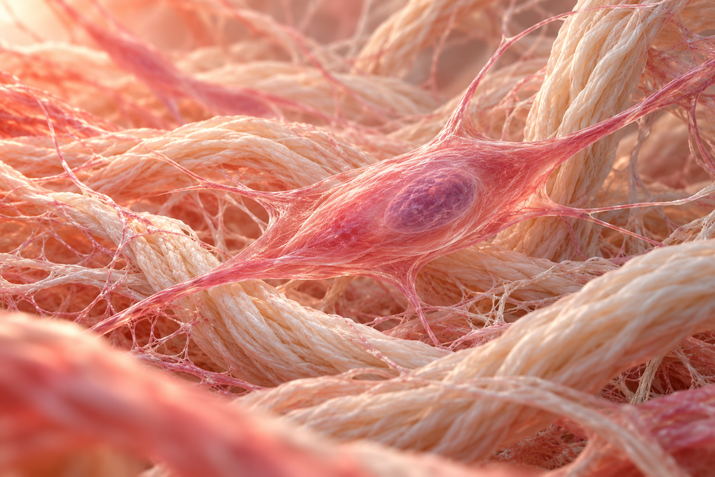
The same experimental study examined AKT, a protein kinase, and the Nrf2/HO-1 pathway. A kinase modifies proteins through phosphorylation. Nrf2 helps regulate cellular defense genes, while HO-1 is an enzyme involved in stress responses.
Researchers connected this signaling with reduced expression of several matrix metalloproteinases, or MMPs, in aged mouse skin.
MMPs participate in normal tissue turnover. The goal is appropriate regulation, rather than eliminating collagen breakdown.
This adds a second dimension to red light therapy and collagen synthesis. Collagen accumulation can reflect changes in both production and degradation. In that mouse model, the researchers investigated both.
What have human studies found?
Human evidence is important because a detailed laboratory mechanism cannot establish the cosmetic results a person will experience.
A 2007 split-face trial enrolled 76 people and compared 633 nm red light, 830 nm near-infrared light, their combination, and sham treatment. Sessions took place twice weekly for four weeks.
Researchers reported improvements in wrinkle and elasticity measurements in the active groups. Tissue examination also showed increased collagen and elastic fibers, alongside activated fibroblasts.
A 2009 investigation combined reconstructed human skin experiments with a separate clinical component. In the skin model, pulsed 660 nm treatment produced a 31% difference in type I procollagen and an 18% reduction in MMP-1 compared with untreated samples.
Those percentages came from the laboratory model. They do not mean participants produced 31% more collagen. The clinical component reported improvements in skin surface and wrinkle assessments.
A 2014 controlled trial reported improved skin roughness and ultrasound measurements interpreted as collagen density after 30 treatments. However, it used gas-discharge lamps, had a nonrandomly recruited untreated control group, and was manufacturer-sponsored.
More recently, a 2025 sham-controlled trial involving 95 women tested a 660 nm mask. Selected wrinkle-length measurements and satisfaction improved, but the groups did not differ significantly on the specialist-rated Wrinkle Assessment Scale.
Together, these studies make red light therapy and collagen synthesis a credible research topic. They also show why the exact measurement matters.
How to read collagen claims without getting lost
Studies can examine different steps in a biological process. Keep these distinctions in mind when reading about red light therapy and collagen synthesis.
| Measurement | What it can tell you | What it cannot establish alone |
|---|---|---|
| Collagen messenger RNA | Whether collagen gene expression changed | How much mature collagen formed |
| Procollagen | Whether a collagen precursor changed | Whether organized fibers increased |
| Tissue staining or microscopy | Changes in sampled tissue structure or collagen-associated signals | The response across every treated area |
| Ultrasound measurements | Changes in acoustic properties interpreted as tissue density | A direct molecular rate of collagen synthesis |
| Wrinkle or elasticity assessments | Changes in a measured skin outcome | Which molecular pathway caused them |
The best explanation connects the measurement to the claim. An attractive before-and-after photograph cannot identify the signaling pathway responsible.
Does more collagen mean fewer wrinkles or tighter skin?
Wrinkles, elasticity, and laxity describe different features. A treatment can improve one measured feature without producing an obvious lifting effect.
The 2007 trial reported changes in wrinkles and elasticity alongside tissue findings. That is a useful connection, but it does not establish that all forms of sagging respond similarly.
Likewise, the mixed outcomes in the 2025 trial show that a favorable result on one assessment may coexist with no significant difference on another.
When considering red light therapy and collagen synthesis, ask what you want to change. Fine surface lines, skin texture, and more pronounced looseness require separate evidence.
“Supports collagen” is too broad to predict the size of a visible result.
Which wavelengths and treatment schedules matter?
The human studies discussed here used specific protocols, including 633 nm and 660 nm red light and 830 nm near-infrared light.
Wavelength describes light’s spectral characteristics. Irradiance describes power delivered per unit area. Fluence describes energy delivered per unit area over an exposure.
Those variables help define treatment. Session duration alone does not.
The 2025 mask trial compared two and three weekly applications over four weeks. More sessions did not significantly improve the reported outcomes under that protocol. That finding does not establish an ideal schedule for every device.
For red light therapy and collagen synthesis, a familiar wavelength printed on a product page is only part of the information you need. Look for research that matches the device, exposure, and intended outcome as closely as possible.
Follow its instructions for distance, session length, and eye protection.
What should you expect from your skincare routine?
The studies discussed here evaluated treatment courses over weeks and repeated sessions. Their results cannot tell you that a single session has visibly rebuilt your skin.
If you track changes, use photographs taken with consistent lighting, distance, angle, and facial expression. Shadows and positioning can change how pronounced lines appear.
The research on red light therapy and collagen synthesis supports measured expectations. It does not provide one guaranteed percentage improvement, universal timetable, or permanent result.
A broader collagen research overview can help you put individual findings in context.
This article is educational and is not medical advice. Consult a qualified clinician about personal skin concerns, photosensitivity, or medications that may affect your suitability for light treatment.

Frequently asked questions
Does red light therapy increase collagen synthesis?
Research on red light therapy and collagen synthesis includes human tissue findings and laboratory measurements consistent with collagen-related effects. The strength of a claim depends on whether researchers measured gene expression, precursors, tissue collagen, or visible skin outcomes.
What do fibroblasts do during treatment?
Fibroblasts maintain the extracellular matrix. Human tissue research has reported signs of activated fibroblasts after particular light protocols. Their involvement is one reason red light therapy and collagen synthesis are studied together, but it does not establish an identical response in every user.
How long does it take to improve wrinkles?
Trials have assessed outcomes after repeated sessions over several weeks or longer. Improvements vary with the protocol and measurement. A study’s assessment date does not establish exactly when an individual first notices a change.
Does red light therapy prevent collagen breakdown?
Experimental research has investigated MMP regulation, including findings in reconstructed skin and mice. This is relevant to red light therapy and collagen synthesis, but it cannot establish that every home device prevents collagen loss in people.
Is red light or near-infrared light better for collagen?
Human studies have tested both, including combined protocols. The evidence discussed here does not establish one universally superior wavelength. Dose, delivery, and the outcome being assessed also matter.
Can it tighten sagging skin?
Evidence of collagen-related activity or improved fine lines does not establish a lifting effect. Red light therapy and collagen synthesis research should be interpreted according to the specific skin changes measured, rather than treated as a promise that loose skin will tighten.